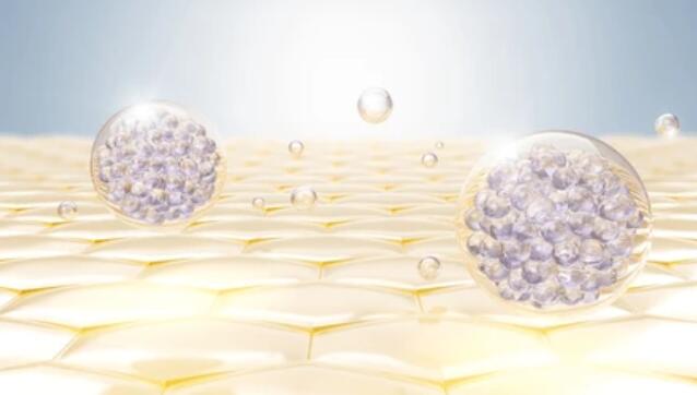
Extracellular vesicles are small membrane-bound particles secreted by cells that are thought to function as intercellular messengers, transporting substances from one cell to another. Extracellular vesicles have several subtypes that vary in function, substance, and size, ranging from about 30-1,000 nm in diameter. One of the smallest types of extracellular vesicles is the exosome.
In biomedical research, exosomes and their carriers are used as diagnostic biomarkers for cancer and other diseases. Exosomes can be isolated from blood or other biological fluids using ultracentrifugation, PEG precipitation, and immunomagnetic beads. The exosome membrane contains transmembrane proteins derived from the plasma membrane of the original cell, usually including proteins of the tetraspanin family (e.g., CD9, CD63, and CD81). Internally, exosomes contain cytoplasmic components such as proteins and RNA, which can be analyzed using RNA-seq, Western blot, and flow cytometry.

Flow cytometry is a fluorescence-based assay that simultaneously determines multiple characteristics, such as cell population counts and protein abundance, from a single cell suspended in solution. It is a powerful tool for rapid, quantitative, and accurate determination of cellular characteristics and provides excellent interpretation against cell population heterogeneity. Flow cytometry allows researchers to use fluorochromes that stain exosomes such as membranes and nucleic acids and antibodies that bind to tetraspanin or other target proteins. Lifeasible offers a range of antibody development services for exosome flow cytometry validation.
| Antibody Name | Clone Name | Notes |
| CD9 (human) | CD9/1619 | Recommended, high brightness of fluorescent coloring. |
| CD9/2343 | ||
| CD81 (human, mouse, rat) | 1.3.3.22 | |
| rC81/3442 | ||
| Rabbit CD81 (human, mouse, rat) | C81/2885R | Slightly weaker than other CD81 antibodies. |
| CD63 (human, mouse) | MX-49.129.5 | Can be used to label unpurified exosomes sorted by magnetic beads. |
| CD63 (human) | LAMP3/968 | |
| Ep-CAM (human) | VU-1D9 | MCF-7 cell-derived exosome shows a low expression of Ep-CAM protein. |
| rVU-1D9 | ||
| EGP40/826+ EGP40/837+ EGP40/1110+ EGP40/1120 |
||
| HEA125 |
We also offer exosome cell membrane fluorochrome stains, which are characterized by higher fluorescence intensity, low background value, removal of non-specific adsorption of the stain without affecting the isoelectric point of the antibody, higher stability with no loss of activity over a long period of irradiation, and higher acid and alkali tolerance.
Lifeasible uses fluorescently labeled surface proteins, membrane lipids, cytosolic esterases, and antibodies targeting cell surface and intracellular markers for simultaneous analysis to gain more information about exosomes. Please feel free to contact us for a customized solution.