The core of the endomembrane system is the endoplasmic reticulum, an essential pleiotropic organelle. With its network of interconnected tubules and flat cisterns, the ER represents the organelle with the largest membrane surface area. It can be viewed as the gatekeeper of secretory pathways that control multiple checkpoints in protein biosynthesis. Morphologically, the ER is able to reorganize, expand, and contract its highly dynamic polygonal tubular network in space and time.
Powered by our professional scientists and their years of field experience, Lifeasible is able to shape the plant endoplasmic reticulum. With our cutting-edge platforms, we can achieve shaping by organelle structures, related gene regulation, and so on.
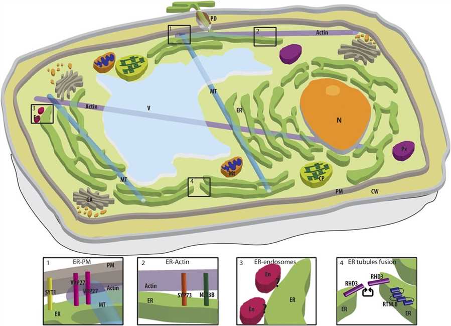
 Fig.1 Association between the ER and other organelles in a plant cell. (Stefano G, et al., 2017)
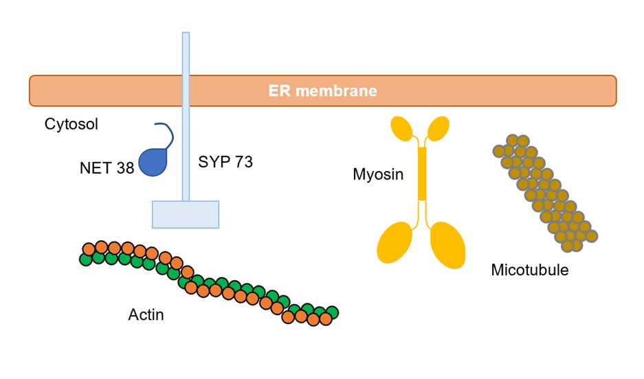
 Fig.2 ER and cytoskeleton interactions.
Lifeasible offers the shaping services involved in the plant endoplasmic reticulum for your research convenience. Our goal is to help our clients achieve meaningful results through powerful and consistent approaches. If you are interested in our services or have any questions, please feel free to contact us or make an online inquiry.
Reference
Lifeasible has established a one-stop service platform for plants. In addition to obtaining customized solutions for plant genetic engineering, customers can also conduct follow-up analysis and research on plants through our analysis platform. The analytical services we provide include but are not limited to the following:
Get Latest Lifeasible News and Updates Directly to Your Inbox
Adaptive Evolutionary Mechanism of Plants
February 28, 2025
Unraveling Cotton Development: Insights from Multi-Omics Studies
February 27, 2025